Eye Exams
Routine Eye Exams are an important part of maintaining good eye health. When its time for your annual check-up, here is some helpful information to get you prepared!

Routine Eye Exams are an important part of maintaining good eye health. When its time for your annual check-up, here is some helpful information to get you prepared!
Part of the examination, such as taking your medical history and the initial eye test, may be performed by a clinical assistant. Several different tests may be performed during the eye exam. The tests are designed to check your vision and to examine the appearance and function of all parts of your eyes.
This test evaluates the muscles that control eye movement. Your eye doctor watches your eye movements as you follow a moving object, such as a pen or small light, with your eyes. He or she looks for muscle weakness, poor control or poor coordination.
This test measures how clearly you see. Your doctor asks you to identify different letters of the alphabet printed on a chart (Snellen chart) or a screen positioned some distance away. The lines of type get smaller as you move down the chart. Each eye is tested separately. Your near vision also may be tested, using a card with letters similar to the distant eye. The card is held at reading distance.
Light waves are bent as they pass through your cornea and lens. If light rays don’t focus perfectly on the back of your eye, you have a “refractive error.” Having a refractive error may mean you need some form of correction, such as glasses, contact lenses or refractive surgery, to see as clearly as possible. Assessment of your refractive error helps your doctor determine a lens prescription that will give you the sharpest, most comfortable vision. The assessment may also determine that you don’t need corrective lenses.
Your doctor may use a computerized refractor to estimate your prescription for glasses or contact lenses. Or he or she may use a technique called retinoscopy. In this procedure, the doctor shines a light into your eye and measures the refractive error by evaluating the movement of the light reflected by your retina back through your pupil.
Your eye doctor usually fine-tunes this refraction assessment by having you look through a mask-like device that contains wheels of different lenses (phoropter). He or she asks you to judge which combination of lenses gives you the sharpest vision.
Visual Field Test (Perimetry)
Your visual field is the full extent of what you can see to the sides without moving your eyes. The visual field test determines whether you have difficulty seeing in any areas of your overall field of vision. The different types of visual field tests include:
Your eye doctor sits directly in front of you and asks you to cover one eye. You look straight ahead and tell the doctor each time you see his or her hand move into view.
A more advanced visual field test when eye disease such as glaucoma is suspected or being treated. As you look at a screen with blinking lights on it, you press a button each time you see a blink.
Using your responses to one or more of these tests, your eye doctor determines the fullness of your field of vision. If you aren’t able to see in certain areas, noting the pattern of your visual field loss may help your eye doctor diagnose your eye condition.
If you have difficulty distinguishing certain colors, your eye doctor may screen your vision for a color deficiency. To do this, your doctor shows you several multicolored dot-pattern tests. If you have no color deficiency, you’ll be able to pick out numbers and shapes from within the dot patterns. If you do have a color deficiency, you’ll find it difficult to see certain patterns within the dots. Your doctor may use other tests, as well.
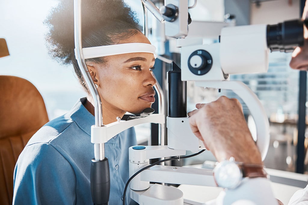
A slit lamp is a microscope that magnifies and illuminates the front of your eye with an intense line of light. Your doctor uses this device to examine the eyelids, lashes, cornea, iris, lens and fluid chamber between your cornea and iris.
Your doctor may use a dye, most commonly fluorescein (flooh-RES-een), to color the film of tears over your eye. This helps reveal any damaged cells on the front of your eye. Your tears wash the dye from the surface of your eye fairly quickly.